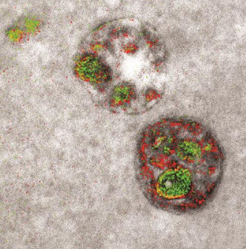
Colors Seen in Images Made From Electron Microscopes Are
So far the researchers can only produce two colorsred and green they report online today in Cell Chemical Biology. Electron microscope images are black and white indicating presence or lack of electrons.

Electron Microscope Captures Cells In Colour For The First Time Research Chemistry World
Electron microscopes can magnify an object up to 10 million times allowing researchers to peer into the inner workings of say a cell or a flys eye but until now theyve only been able to see in black and white.

. Elsewhere winter is giving. Standard EM images are in grayscale and any color is added in with computer graphics programs after the image is made. Still the ability to use color creates stark contrasts that grayscale images simply cant accomplish.
So what you are looking at the image produced by an electron microscope is basically the contrast. The microscope detects when each metal loses electrons and records each unique loss as an artificial color. Bringing color to electron microscope images is a tricky problem.
Scanning electron microscopy SEM in particular has given us some striking images over the years to tantalize our visual senses. Finally a light microscope allows you to see the specimen exactly how it is meaning in full color. A research team from the University of California San Diego is the first to create a multicolor electron microscope allowing for three colors at a time green red and yellow.
You dont know snow unless youve seen it really close up. The colored images from SEM or TEM are manipulated and colored after they acquired. Colors seen in images made from electron microscopes are Added to make certain structures easier to see Which type of microscope can produce three dimensional images of a cells surface.
In some regions of the US spring is upon us. The ability of these microscopes to help us visualize specimens. Image b is the height map used to produce a 3D mesh.
Thats kind of what it was like for the scientists who have taken the first multicolor images of cells using an electron microscope. However with an electron microscope you can view it in 3D. Click here to get an answer to your question Colors seen in images made from electron microscopes are rizzy26 rizzy26 03092017 Biology High School answered Colors seen in images made from electron microscopes are 1 See answer.
Electron microscopes offer us the capability to study things right down to the atomic level. The area where electrons pass through the specimen appears white and the area where electrons do not pass through appears black. The team could see a string of proteins.
The rainbow color scale applied here is traditionally used to express height on a physical map where the lowest points are blue the sea the plains are green the mountains are red and the highest points are white snow-capped peaks. And again - the origan maps are BW. The Ebola virus itself is too small to be seen even in a high-powered light microscope.
Colors seen in images made from electron microscopes are what. SEM involves scanning a focused beam of. In other words we can look at single.
Osmium tetroxide OsO 4 is a popular stain for TEMproviding the characteristic black color and contrast within the sample. Colors seen in images made from electron microscopes are a. 14 Striking Photos of Snow Under an Electron Microscope.
The exception is with stereo microscopes which uses two eyepieces to create a 3D image. Adams et alCell Chemical Biology 2016 Because electron detectors work this way the end product of an electron microscope is. See the first color images produced by an electron microscope.
The colors of electrons c. The pattern of how the electrons pass through or are deflected produces the image. It could plausibly be said that color doesnt exist at that scale because the things imaged by an electron microscope are.
Electron microscopy EM is a time-honored technique for visualizing cell structures that uses beams of accelerated electrons to magnify objects up to 10 million times their actual size. With an electron microscope the image is seen in black and white. An electron microscope is a highly advanced microscope that depending on the type of electron microscope blasts electrons through a specimen excites electrons that make up the specimen or maps the tunneling of electrons through a specimen and reconstructs the feedback from these methods to form an image.
True to life b. Electron microscopes can magnify an object up to 10 million times allowing researchers to peer into the inner workings of say a cell or a flys eye but until now theyve only been able to see in black and white. An electron microscope is a sophisticated and versatile imaging device that can be used to study live and fixed organic and inorganic materials as well as observe their transformation over a certain period of time.
Added to make certain structures easier to see. Added so scientists can trace living cells through the body. EDSEELSEF-TEM maps can have different colors - every color will represent different element.
But when visualized in an electron microscope the surface of the filament-shaped virus particle also known. In the new color TEM technique rare earth metals lanthanides are used to produce the different colors. Each color highlights a different component of the cellular ultrastructure Normally electron microscopy labels structures with gold particles but they tend to show up as nondescript black.
The colors were generated using a new electron microscope technique.

This Is A False Color Image Of A Transmission Electron Micrograph Of A Synapse The Bright Pink Areas Microscopic Photography Cells Project Patterns In Nature

Beautiful Partially Colored Electron Microscopy Of A Cell Nucleus Nuclear Envelope And So On Biologia Celular Biologia Microscopio

Ldeo On Twitter Things Under A Microscope Electron Microscope Electron Microscope Images

Comments
Post a Comment